3D Mapping of Viruses by Electron Microscopy
Understanding the characteristics of a pathogen is a prerequisite for developing safe and efficacious vaccines and medicines. Thus, it is known that the surface protein of the SARS-CoV-2 pathogen – also called spike protein – plays a key role in the virus replication cycle.

The spike protein mediates the cell binding and the fusion of virus and cell membrane and this initiates the infection of a cell. In addition, the spike protein determines the host range and cell tropism. Cryo-electron microscopy (cryo-EM) enables to resolve virus structures in detail, at a resolution close to an atomic scale. That way, the structure of the spike exposed on the surface of SARS-CoV-2 can be resolved and compared to that of other coronaviruses. At the Paul-Ehrlich-Institut, Federal Institute for Vaccines and Biomedicines, the research group "Electron Microscopy of Pathogens", headed by Professor Jacomine Krijnse Locker, applies 3D cryo-EM for the analysis of viruses such as SARS-CoV-2.
The focus of the research group is the establishment of modern methods in EM to obtain high-resolution images of viruses. As their model, they use large DNA viruses. Among other things, the research group was able to show that DNA viruses break up membranes of the infected cells for usage in the biogenesis of the virus envelope. In collaboration with researchers of the university hospital of Heidelberg, the research group used EM to obtain high-resolution images of Dengue virus- and HIV-induced modifications of cellular membranes. Thus, 3D-EM revealed that for flaviviruses, the replication of the virus genome and the virus budding are highly co-ordinated, which facilitates the formation of infectious particles.
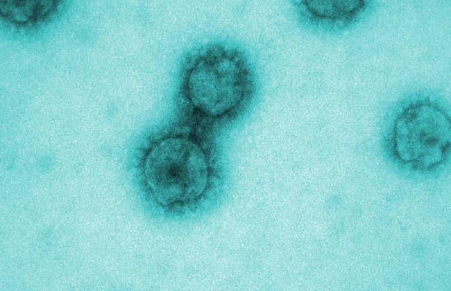
Since the outbreak of the SARS-CoV-2 pandemic, the group has worked on this novel coronavirus. First images of the new pathogen have already been generated and show pleomorphic virus particles (particles with different shapes), which carry the typical spikes on their surface. The aim is to achieve a detailed high-resolution characterisation of the viral structures by means of cryo-EM to understand why this virus spreads so successfully. These findings could provide information on how the virus structures can be modified to inactivate the virus and block infection.
Reinforcing the Research by Imaging Methods at the Paul-Ehrlich-Institut
Jacomine Krijnse Locker obtained her PhD at the University of Utrecht (the Netherlands). After that, she spent several years working at the European Molecular Biology Laboratory (EMBL) in Heidelberg and at the University of Heidelberg. Then she was the head of the Electron Microscopy Group at the renowned Institut Pasteur in Paris. Here, she performed research on the mechanisms underlying the entry of the HIV genome into the host cell nucleus. In 2019, Professor Krijnse Locker followed the appointment to the Justus-Liebig University at Gießen as a Loewe-DRUID professor for "Neglected Diseases and Imaging Methods". Since January 2020, she has been head of the research group "Electron Microscopy of Pathogens" at the Paul-Ehrlich-Institut.


